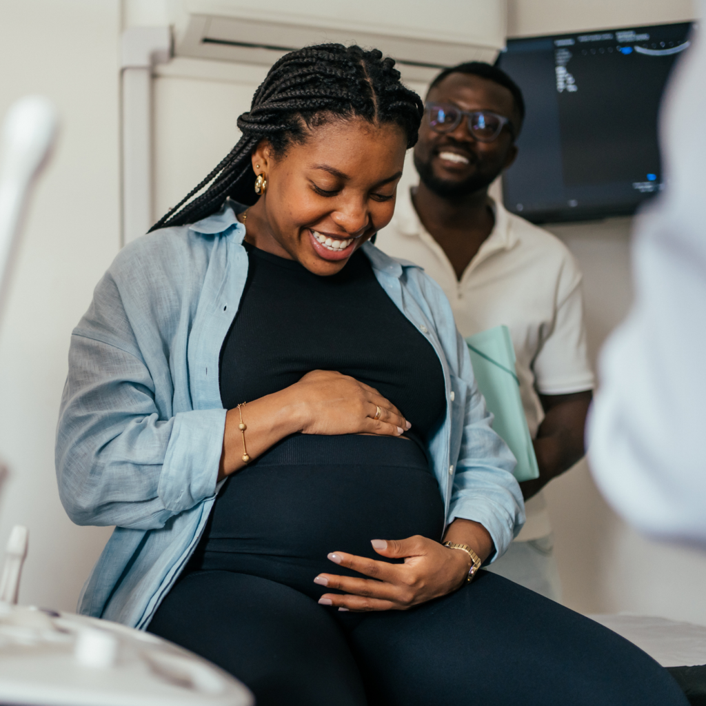
Advances in prenatal diagnosis and fetal surgery are changing what’s possible before a child is even born. Three specialists from the Institute for Maternal Fetal Health at Nemours Children’s Health discuss how breakthroughs in fetal care are improving outcomes for babies with complex conditions. They also share how a multidisciplinary team supports families through prenatal diagnoses, difficult decisions and every step of the journey – from pregnancy to delivery and beyond.
Featuring:
Julie S. Moldenhauer, MD, PMH-C, FACOG, FACMGG, Executive Director, Institute for Maternal Fetal Health, Elizabeth W. Snyder Endowed Chair in Fetal Therapy and Innovation, Nemours Children’s Health
Eric Paul Bergh, MD, Director of Fetal Research and Informatics, Institute for Maternal Fetal Health, Nemours Children’s Health
Christina (Nina) Paidas Teefey, MD, PMH-C, Director of Psychosocial Programs, Institute for Maternal Fetal Health, Nemours Children’s Health
Host/Producer: Carol Vassar
TRANSCRIPT
Announcer (00:00):
Welcome to Well Beyond Medicine, the world’s top-ranked children’s health podcast produced by Nemours Children’s Health. Subscribe on any platform at nemourswellbeyond.org or find us on YouTube.
Carol Vassar, podcast host/co-producer (00:12):
Each week we’ll be joined by innovators and experts from around the world, exploring anything and everything related to the 85% of child health impacts that occur outside the doctor’s office. I’m your host, Carol Vassar. And now that you are here, let’s go.
MUSIC (00:30):
Let’s go, oh, oh, well beyond medicine.
Carol Vassar, podcast host/co-producer r (00:36):
You know, every day, parents looking forward to the birth of a child walk into what they think is an ultrasound that is routine, and they’re expecting reassurance and hoping for the healthy development of their unborn baby. Occasionally, though, that is not how the scenario goes, and a routine scan reveals a serious diagnosis that in some cases requires immediate pre-birth intervention, often in the form of surgery.
(01:04):
In this latest episode of an occasional series on maternal fetal health, we’re going to explore how advances in prenatal diagnosis, fetal surgery, and integrated care are reshaping what’s possible before a child is even born to treat complicated diagnoses. Joining me right now from the Institute for Maternal Fetal Health at Nemours Children’s Health are its executive director, Dr. Julie Moldenhauer, along with Dr. Eric Bergh. He’s a fetal surgeon and director of fetal research and informatics. And Dr. Christina Nina Paidas. She’s a fetal surgeon and director of psychosocial programs within the institute.
(01:45):
Dr. Moldenhauer, I’m going to go to you first. Let’s take that ultrasound example for a moment and start with prenatal diagnosis. Talk about the importance of getting prenatal care in general, and tell us what happens. At what point in the process does your team get involved?
Julie Moldenhauer, MD, Nemours Children’s Health (02:04):
Yeah. So I mean, as we all know, healthy pregnancies really require sound prenatal care, which also requires access to sound prenatal care for women once they learn that they are pregnant. And so there are many points during the pregnancy where patients will present not only for an office visit for, say, like a blood pressure check or a prenatal visit, but then an imaging visit. And so some of these imaging visits, they can happen very, very early in the pregnancy. And we call those dating ultrasounds where we’re really just kind of confirming: is there a singleton? Are there multiples? Are there twins or triplets? Does everybody have a normal heartbeat? And what is the presumed due date based on measurements of a very early ultrasound? Because that’s the most accurate form of dating that’s available to us.
(02:51):
And then, towards the end of the first trimester, around 12 to 14 weeks, we can also do a limited anatomy ultrasound. And so some of the more major anomalies or differences can be identified very early in the pregnancy, given with how far along we’ve come with our imaging technology, which is humongously important. Because if we can identify a difference very early, we can make referrals to a fetal care center or a higher level of care available for the patient.
(03:21):
And then what we would consider sort of the routine anatomy ultrasound, where we really are looking at fingers and toes and all the details of the heart and the brain and so on and so forth, that really happens in the second trimester, so between about 18 and 22 weeks of the pregnancy. But at any point in time, we could find a difference that would prompt a referral to a fetal care center for the next level higher of evaluation that might encompass not only a higher level, more detailed ultrasound, but also a fetal echocardiogram where we’re getting really good views of the fetal heart or even a fetal MRI where we’re looking at just much finer detail, say, of brain structures or lung structures or something like that.
Carol Vassar, podcast host/co-producer (04:09):
Let’s move ahead to that moment when you do find an anomaly. And I know you’ve all had this experience, but Dr. Paidas, I’m going to go to you. How do you break that kind of news to a family? It’s really going to upend their lives, if only temporarily.
Christina (Nina) Paidas, MD, Nemours Children’s Health (04:24):
I think that you do it honestly and with empathy. And I think that that’s hard when you’re under a time limit because you’re scheduled in a certain way, not necessarily in a fetal center, but in a routine MFM office where you have to break that news with compassion, with empathy, and with honesty in 10 minutes, right? And you are actually shattering someone’s dreams and changing their life and changing significantly their day.
(04:53):
So I think that is a hard question. It’s not just an easy one. We all want to do it with our whole heart. We want to do it with sound imaging and with every piece of information that we can give. But often knowledge is not necessarily power and knowledge doesn’t necessarily help the family, but actually increases their anxiety and increases their fear because there’s uncertainty. Like Dr. Moldenhauer said, there’s a need for more imaging or clarification pieces. And that’s one role where the fetal center can help in the setting of a congenital anomaly or a difference when there is concern.
(05:36):
And I do think it’s really important that maternal fetal medicine doctors, of which we are part of that group, are essential to this process. They do an amazing job at these really early ultrasounds before they get to us. And I think we all, as a community, want to deliver that different news, that change, that care with the best possible outcome, the best possible prognosis. And that’s not always necessarily how the day is built for us or the families. So I think that it requires a lot of thought and a lot of efficiency, which grief is not efficient. And so that’s a challenge, too.
Carol Vassar, podcast host/co-producer (06:22):
So very carefully with a lot of heart and a lot of, it sounds like, support, even though the family might be overwhelmed in that moment, their anxiety is going up, your team is there along with all of the teams that work at the Maternal Fetal Health Institute at Nemours to help support them.
(06:40):
Dr. Bergh, I want to ask the question. We’re kind of concentrating on fetal surgery today. What kinds of diagnoses would require fetal surgery? And what is fetal surgery? It sounds kind of intricate.
Eric Paul Bergh, MD, Nemours Children’s Health (06:57):
So there are actually, I’d say, a list of different diagnoses for which there are some interventions that can be performed in pregnancy. The majority of the interventions that we perform are typically what we call lifesaving, although some of the interventions we perform also are designed to improve what we call morbidity or challenges that a baby might face after birth. Some of the interventions we perform are what we call minimally invasive. Those are interventions that can be performed with just a simple needle and an ultrasound. Other interventions require a slightly larger incision and the use of very, very small cameras and instruments to go inside the uterine cavity where the baby is located. And then there are some interventions that require much more invasive surgery, with typically an incision on mom’s abdomen and even an incision on the uterus.
(07:54):
So the types of interventions we perform are performed not only in what we call singleton, just one baby pregnancies, but we also perform interventions in pregnancies where there’s more than one baby. And some of the things that we do are very, very rare and require a really sophisticated team.
(08:17):
So for instance, something like treatment of twin-twin transfusion syndrome is a pretty common surgery that we’re all really trained in. And that’s a surgery where we use very minimally invasive techniques to separate babies that are joined via one placenta. And that’s actually a lifesaving procedure. That’s something that, without that procedure, the vast majority of babies that have advanced twin-twin transfusion syndrome may not survive. And with this procedure, survival rates are extraordinarily high. So it’s something that’s relegated to really centers of excellence, centers with very specific expertise, and we’re really proud to be a center that can offer those kinds of interventions.
Carol Vassar, podcast host/co-producer (09:05):
Dr. Moldenhauer, I’m curious, you mentioned the ultrasounds that happen throughout the early trimesters, the first two trimesters of a pregnancy. Is there a point where you just say, “We’re going to watch this”? And is that a possibility? And also, at what point do you say, “Hey, we need to consider surgery”?
Julie Moldenhauer, MD, Nemours Children’s Health (09:28):
That’s a great question. So many of the conditions we really just expectantly manage. And to get a good outcome for mom and baby just involves a lot of monitoring, close surveillance, and a lot of follow-up. And so the overwhelming majority of patients that we see aren’t actually going to need a fetal intervention in utero. It’s monitoring. But for a certain number of conditions, we know what the natural history of those disorders are. And there are almost guidelines or criteria that we use to help guide us in this scenario.
(10:03):
So for example, Dr. Bergh mentioned twin-twin transfusion syndrome. Not every set of monochorionic twins that has some pre-aspects or some of the markings of twin-twin transfusion syndrome would be candidates to have the laser therapy. For example, it may be sort of just discordant amniotic fluid, and they haven’t met the firm criteria. Or if it’s stage one twin-twin and there are no other symptoms or a short cervix or any other findings, we can just continue to observe those until they have crossed the threshold to a stage two or higher where definitely we know they would need therapy.
(10:40):
For some other disorders, it’s more of an elective type of procedure. So for example, closure of spina bifida in utero, that really is more of an elective type of procedure where parents may want to follow that pathway because they want to try to improve the quality of life for the newborn and the child longer term.
(11:02):
And then there are other conditions. For example, if a fetus presents itself in heart failure, what we call high drops, doing a procedure, a surgery, or intervention may be more urgent. Because if we don’t do anything right away, the baby could pass away well before birth. And so a lot of it just depends on what the underlying diagnosis is, and then what are all the elements around that diagnosis based on what we know of the natural history for each diagnosis. And this is where, when you come to a fetal center, you’re going to see experts who have taken care of many different cases with all of these rare diagnoses. And so we know when we’ve crossed that threshold and an intervention would really be required. Sometimes that intervention could be an earlier or a preterm delivery as well.
Carol Vassar, podcast host/co-producer (11:52):
I was going to ask about that. Dr. Paidas, what are some of the potential outcomes with fetal surgeries, risks in particular to both mother and child?
Christina (Nina) Paidas, MD, Nemours Children’s Health (12:02):
Yeah, so no matter how you look at it, you’re taking care of more than one person. And so we have to be thinking not only of one individual, but we have to think of a family. And we’re not just thinking of them in the sense of thinking of your family of four sometimes. We’re thinking of them all living in the same house, in the same room, all using the same bathroom and the same pantry for their food kind of thing.
(12:28):
And so when we think about risk, it’s a really important discussion. And going back to what Dr. Moldenhauer said, that’s why a lot of the time the risk-benefit ratio favors watching and not necessarily doing. And it’s hard sometimes to keep your hands tied and not just go in to do something when it’s not yet the right time. So there’s risk to the mother, the pregnant person, and there’s risk to the baby or babies, the fetuses on the inside of the uterus.
(12:59):
And those risks can range from something as simple as a little dip in the heart rate that comes right back up with a fetal surgery to separation of the membranes. I call it the expensive wallpaper in the uterus that actually kind of… If it’s not glued the way it’s supposed to be glued, or if somebody comes through it with an instrument or something, it interrupts the natural process and physiologic nature of those membranes. And if that membrane separation or the separation of the wallpaper, the glue doesn’t hold, that can increase your risk for breaking your water early and having an early delivery. We’re always thinking about early delivery. We’re always thinking about implications of early delivery.
(13:41):
But I think that the other risk, honestly, is the risk to the mom. The risk to the mom without doing a procedure. And that is a possibility when a baby has a condition like heart failure, or we call high drops in utero. That actually can result in sequelae in the mother where there’s something called mirror syndrome, where the mother mirrors the sickness of the baby with a hypertensive disease; that can actually cause organ damage and be life-limiting for the mother. So there can be risk on the inside without doing a procedure, and there also can be risk with the procedure. And I think that those are some of the things that we think about, but there’s always, actually, the worst possible outcome, too, for the family that may be dying, the baby dying, or actually sometimes life-altering life, which can be equally as hard as dying.
Carol Vassar, podcast host/co-producer (14:45):
As with any surgery, whether you’re an adult or a child or in utero, there are always risks. I want to continue the conversation, Dr. Bergh, and talk about how you shape the conversations with families that you have about those decisions that are made, especially with the mother and child in mind.
Eric Paul Bergh, MD, Nemours Children’s Health (15:05):
Well, I think any conversation begins with a really accurate description of what’s going on. I like to think of myself and my colleagues as educators first, and we’re really there to help walk a patient through what is a very complex diagnosis, and typically a diagnosis they’ve just learned about, so that they can make what is what we would hope an informed decision, as informed as they can be, and laying out, just as we’ve just spoken about, what are the risks associated with doing nothing versus the risks that we might encounter if we try to be more aggressive and do something like a fetal intervention.
(15:47):
And for the most part, we are just watching and waiting. But when it comes time that we actually have to do an intervention, the most important thing is that the patient, the patient’s family, is fully aware of what we’re moving forward with, because it changes the course of the entire pregnancy sometimes. And so my priority is making sure that a family is fully informed so that when they do ultimately consent for any decision that they make, they feel like they are making that decision with all of the possible knowledge that they need to make that decision.
(16:22):
I think one of the other really important parts of the conversation is trying to give them the best possible idea of what life looks like after pregnancy. And sometimes that involves not only pulling from my own resources, but also pulling from colleagues in other specialties. And we work really closely together with other pediatric subspecialists who can help us round out some of the discussion as to what things are going to look like in pregnancy, but also: what does life look like after pregnancy. And I think that’s part of doing the best job we can to inform our families.
Carol Vassar, podcast host/co-producer (17:04):
And I do want to talk about that multidisciplinary approach shortly, but I want to open a question up to whichever one of you would love to respond to this because I would like to know if there’s a patient story that really illustrates what’s possible through fetal intervention, what was the diagnosis, without going into anything that’s HIPAA, not revealing anything about the family specifically, the choices they faced, and maybe even where that child is today, how they’re doing.
Eric Paul Bergh, MD, Nemours Children’s Health (17:36):
Yes. So I had the privilege of taking care of a family that was facing a challenging diagnosis in which they were referred to our facility for evaluation of a fetus that, on ultrasound, had fluid inside the chest. So more accurately, there was fluid in the space where the lungs are on one side of the chest, in the space between the chest wall and the lung. So the technical term for this is a pleural effusion, and a pleural effusion can occur for a lot of different reasons.
(18:15):
But ultimately, what was going on, I think, really, we determined that it was a lymphatic issue, that some babies, their lymphatic system develops a little bit more slowly. And so rather than the lymph fluid draining, it can accumulate in spaces around the body. And if you get a large fluid accumulation in the chest, that can actually cause some problems for the baby in utero. It pushes on the heart; it can cause the heart to work extra hard, and it can cause the baby to go into a little bit of what we call heart failure.
(18:47):
Or sometimes that effusion might be small, like a small amount of fluid, and we typically watch that, like we’ve alluded to here. We don’t always jump into intervention right away. We watch to see what is the evolution of that, because many pleural effusions will regress or go away on their own. But occasionally they may be more aggressive. They may grow. And in this case, this is a case in which the effusion continued to expand, expand in such a way that we identified that the baby was at risk for developing heart failure and was showing early signs of distress.
(19:21):
So the typical workup or the way we proceed there is a discussion about maybe doing an intervention to try and reduce the amount of fluid in the chest, ultimately to help stabilize baby and let the pregnancy continue on, to try to avoid a preterm birth or an adverse outcome. And so we did do a procedure. We started with a very minimally invasive needle-based procedure where we used an ultrasound to guide a very long, thin needle into the baby’s chest, and we withdrew fluid from the chest, letting that healthy lung expand into the space. And we observed to see how things evolved over time. And what we saw over the course of several days is that the effusion returned very aggressively.
(20:07):
And we’d been hopeful that maybe just one procedure would solve the problem, but because it didn’t, the next step was a discussion about placing a shunt. So a shunt is a small plastic catheter that we can use to essentially connect the space inside the chest with the amniotic space. And using a very small needle relative to what we normally do in fetal surgery, we can place that shunt with the needle into the baby’s chest so that it crosses or it spans the fetal chest. And so one part of the shunt will be in the space where the fluid is, and the other part of the shunt will be outside the baby. And by placing that shunt, we let the fluid that accumulates in the chest just kind of drain out of the chest into the space around the baby.
(21:00):
And we were really lucky to be able to offer that procedure to this patient. We did a minimally invasive procedure in the operating room using ultrasound where we were able to accurately place that shunt into the correct position such that within 24 hours, all the fluid had drained out and it stayed out throughout the remainder of the pregnancy. So any fluid that the baby made, it just kind of drained out, and the heart went back to its normal position. And all the things that we were worried about just kind of reversed.
(21:29):
And that baby did really wonderfully. That baby continued on in utero for weeks until the baby got to full term and had a really uncomplicated delivery. And postnatally, the shunt was removed, and baby did really, really well. We’ve seen that baby now a couple of times and is really what we say thriving. And I think the wonderful part of the story here is that we were able to progressively work this up, figure out what was needed when it was needed, and we were able to avoid a preterm birth, and we were able to avoid a really sick kid, and ultimately have a really wonderful outcome for this family.
Carol Vassar, podcast host/co-producer (22:10):
That is fabulous news, and congratulations on that. It’s an outcome that we all hope for. As I’m listening to all of you talk, I’m hearing that technology plays a big role in how you do this. When I look back at when I was pregnant the first, second, third time, 30, 33, and 35 years ago, these kinds of interventions, to the best of my knowledge, were not available. Dr. Moldenhauer, I’m wondering how the development of technology has made it possible for these kinds of interventions, these kinds of surgeries to take place.
Julie Moldenhauer, MD, Nemours Children’s Health (22:50):
I mean, clearly, imaging – the evolution of imaging capabilities in the prenatal diagnostic space has been front and center of what we’ve been able to do. Ultrasound capabilities, in particular, have just come light years in the last 20 to 30 years. The imaging is so crisp now. You can really make out structures in the baby where it used to be a little bit what we would call grainy or difficult to really envision or separate different parts of the fetus on ultrasound.
(23:25):
So when you look at an ultrasound, anything that’s fluid is black. Anything that’s very dense, like bone, is bright white. And all of the soft tissues are sort of shades of gray in between. We’re really able now to sort out all those different shades of gray so that we can look at kidneys and say, “Wow. Those are totally normal kidneys. That’s a totally normal adrenal gland,” or “That’s a totally normal-looking liver,” or spleen or bowel or those sorts of things.
(23:50):
And even just the various structures inside the brain that we can make out very distinctly. The corpus callosum, we can now see that very well. It’s a midline structure, and we can find the blood vessels that feed that midline structure in the brain that 15, 20 years ago was very hard to make that out unless you did an MRI. And so first and foremost, our imaging capabilities have just gone above and beyond.
(24:18):
And then our ability with technology as far as the instrumentation that we use in the operating room, everything is getting much smaller, much more refined, so that the damage that we’re potentially doing to the uterus or the membranes more so becomes less and less. And so a lot of the complications that we would see that Dr. Paidas and Dr. Bergh have mentioned with the membrane separation or rupturing the membranes, those sorts of things is becoming less and less because we’re able to do a lot more through very fine, smaller ports or incisions on the uterus.
(24:56):
Some of the surgeries that we initially would do through those very large incisions on mom’s abdomen and on the uterus, we can now do through fetoscopic approaches because we’re able to use instrumentation that’s much smaller and more refined than we’d previously used. And there are certainly many conditions now where we don’t even have to put an instrument in the uterus. We can deliver a medication transplacentally. Mom takes the medication, and then it gets delivered through the placenta to the fetus. Or we can inject the medication into the amniotic sac, and the baby swallows it just as if they were going to take a pill themselves. It gets dissolved in the fluid. And so our knowledge, our technology has just evolved phenomenally so that we’re in a whole nother world than we were 20 years ago.
Carol Vassar, podcast host/co-producer (25:49):
I want to get back to that multidisciplinary aspect of the work you do. Fetal surgery is one piece of the puzzle. What has to happen behind the scenes to move a family from, say, diagnosis to intervention or maybe watching to delivery in ongoing care? Dr. Paidas?
Christina (Nina) Paidas, MD, Nemours Children’s Health (26:10):
Yeah, it takes a village. And even in the operating room with a fetal surgery, fetal intervention, any fetal therapy, it still takes a village. So the village can look different for every family. The village is actually designed for that family. That’s the advantage of a fetal center. And so oftentimes it involves maternal-fetal specialists, including the team from home where the family is being referred from. Of course, nursing and sonographers, imaging techs are a critical part of the intervention team, and it takes years and years of training for them to get to that level of mastery.
(26:57):
The advanced practitioners in women’s health are also trained in the fetal surgery environment, and so we’re also trained to look for and know what prenatal care aspects are most important or critical in this family or in this patient. They know when we see something as a reflex, what they need to look for in the mother.
(27:19):
And then I also think that the genetics team, the genetic counselors, prenatal genetic counselors, and geneticists like… Dr. Moldenhauer wears multiple hats, as you know, is also a geneticist. Having that on deck as a part of your village is a really critical aspect of care. But then you have reach in terms of mental health, psychosocial support, social work, coordination of care, behavioral health support.
(27:50):
And then so far even further, you look toward what the baby may need to know… or what the parents may need to know about the baby on the inside. That’s our team. But also, what does it look like, like Eric said, on the outside? And so you incorporate, and you really rely very much on the expertise of your partners in subspecialty areas. And so that can be our colleagues in neonatology. That can be palliative care specialists. That can be cardiothoracic surgeons or fetal cardiology who span fetal and neonatal care of the baby’s heart. Neurology and neurosurgery are very, very often our partners. Pediatric surgeons that can also do fetal surgery and postnatal surgery that participate in airway delivery, specialty surgeries. Today, ENT was involved in it in a case that we were helping with.
(28:50):
So I think that really, your village is built for your family, your family’s goals. And all of the family really is the center, the sun, the moon, and the stars. Everything revolves around that family, and you build what they need. And you’re just fortunate to be in a center where we have that accessibility, where we have those imaging specialists to walk down the hallway and go look at an MRI with a professional that really values your expertise, but also that you’re in it for that family and you’re both eyes on that baby, on that family to give them the most possible information.
Carol Vassar, podcast host/co-producer (29:30):
I’m glad you brought that up. The Institute for Maternal Fetal Health at Nemours provides continuity from the time of referral, probably beyond. Dr. Bergh, talk about why that continuity is so important.
Eric Paul Bergh, MD, Nemours Children’s Health (29:46):
So our families are dealing with very complicated diagnoses. Not only do their pregnancies look more complicated than a typical pregnancy, but everything that happens afterward is typically more complicated than a normal well-baby stay. And having as many people involved who know the case consistently throughout the pregnancy helps make sure that the messaging is the same from the time of diagnosis until after birth.
(30:20):
I think that one of the main benefits of having a center where we are so intimately involved in the patient’s care is that week to week or month to month, the patients are really seeing the same team members who are intimately aware of all the details of their complicated pregnancy so that the opportunity to miss something is significantly reduced because the same people are so involved from the very beginning to the very end of their time here at Nemours and at the institute. There’s nothing worse than coming into a prenatal appointment and meeting someone new every single time. And we have the luxury of being able to schedule our patients in such a way that they feel like we’re there with them every step of the way.
Carol Vassar, podcast host/co-producer (31:12):
Dr. Moldenhauer, anything to add to that?
Julie Moldenhauer, MD, Nemours Children’s Health (31:16):
Yeah, no. I mean, I think the fact that we get to really walk alongside patients through this very complicated journey is humongously important. And they have the support. They have the support from the medical staff, and then they have the support from Dr. Paidas’s psychosocial team. But additionally, the team that’s going to take care of their newborn in the NICU or the cardiac intensive care unit, they also get the opportunity to be exposed to them, meet them, get to know that team so that when they transition from this prenatal world to the newborn world, they have already met the doctors, many times the nurses, the nurse practitioners. Many aspects of the process in the stay have already been explained to them. So a lot of that mystery or that scary sensation about what’s going to happen when my baby’s born, they’ve at least heard some of it. They’ve taken a tour of the NICU. They’ve taken a tour of the CICU.
(32:08):
So hopefully the whole goal is that, like Dr. Bergh mentioned, that we’re being educators. We’re educating them on every step of the process and hopefully taking some of that really scary edge out of the process because they have enough to worry about with a baby that’s going to be sick.
Carol Vassar, podcast host/co-producer (32:24):
We’ve been talking a lot about procedures, interventions, surgeries that, as we noted earlier, may have sounded impossible a generation ago. Dr. Moldenhauer, final question. What are you looking ahead to, say 10 years down the line in terms of fetal medicine advances? What are you most excited about?
Julie Moldenhauer, MD, Nemours Children’s Health (32:46):
Ooh. I am very excited about what we are going to be capable of doing from a very minimally invasive standpoint where we’re reducing a lot of the maternal risk and the pregnancy risk, but still getting the same outcomes for the babies longer term. And one of those really big areas, at least from a genetic standpoint, is for disorders that we’ve never had any type of cure for beforehand, so like metabolic disorders, glycogen storage diseases, even sickle cell disease. By using gene editing, gene therapy, enzyme replacement therapies, many of these types of therapies that are much more minimally invasive and for some very rare conditions, it’s becoming a reality that we’re going to be able to offer these to families and give them hope for longevity for these complicated and impacted children.
Carol Vassar, podcast host/co-producer (33:45):
Fetal surgeon Dr. Julie Moldenhauer is Executive Director of the Institute for Maternal-Fetal Health at Nemours Children’s Health. We also heard from two additional fetal surgeons from the institute, Dr. Eric Berg, director of fetal research and informatics, and Dr. Christina – Nina – Paidas, who serves as the institute’s director of psychosocial programs.
MUSIC:
Well Beyond Medicine
Carol Vassar, podcast host/co-producer
Thanks so much to Dr. Moldenhauer, Dr. Berg, and Dr. Paidas for joining us and sharing these amazing advances in not just prenatal diagnosis but also fetal intervention, fetal surgery, and coordinated care, all reshaping what’s possible before a child is even born. Amazing stuff. More amazing stuff is coming your way on the Nemours Well Beyond Medicine podcast as next time we learn how Dr. Lisa Fitzpatrick from Grapevine Health is grabbing a camera and a mic and taking health literacy to the streets, literally. Please join us.
Our website is filled with great information, along with all of our past podcast episodes. Check it out at nemourswellbeyond.org. That’s also where you can leave a podcast review or episode idea, and subscribe to the podcast and our monthly e-newsletter. Again, that’s nemourswellbeyond.org. You can also subscribe to the podcast on your favorite podcast app and on the Nemours YouTube page.
Our production team for this episode includes Lauren Teta, Cheryl Munn, Susan Masucci, and Alex Wall. Video production by Josh Hansbrough. Audio production by me. I’m Carol Vassar. Thanks so much for listening. Until next time, remember, together we can change children’s health for good, well beyond medicine.
MUSIC (34:24):
Let’s go, oh, oh, well beyond medicine.